Diagram Of Liver Cell / : 2.3.1 draw and label a diagram of the ultrastructure of a liver cell as an example of an animal cell.. Terms in this set (9). | human cell structure, animal cell project, animal cell. Control of liver cell fate decision by a gradient of tgf beta signaling modulated by onecut transcription factors. It should be large, clear and with specific labels. These then join with veins from other lobules, forming.
Blood drains from the sinusoids into central or centrilobular veins. Expression of liver specific proteins decreases with time in culture, but is reactivated by growing the cells in serum free medium. Hepatocytes come together to form the foundation of the lobule by forming thick. Ƽ intricately involved in carbohydrate, fat, and protein metabolism. Human liver and hepatic cell diagram.
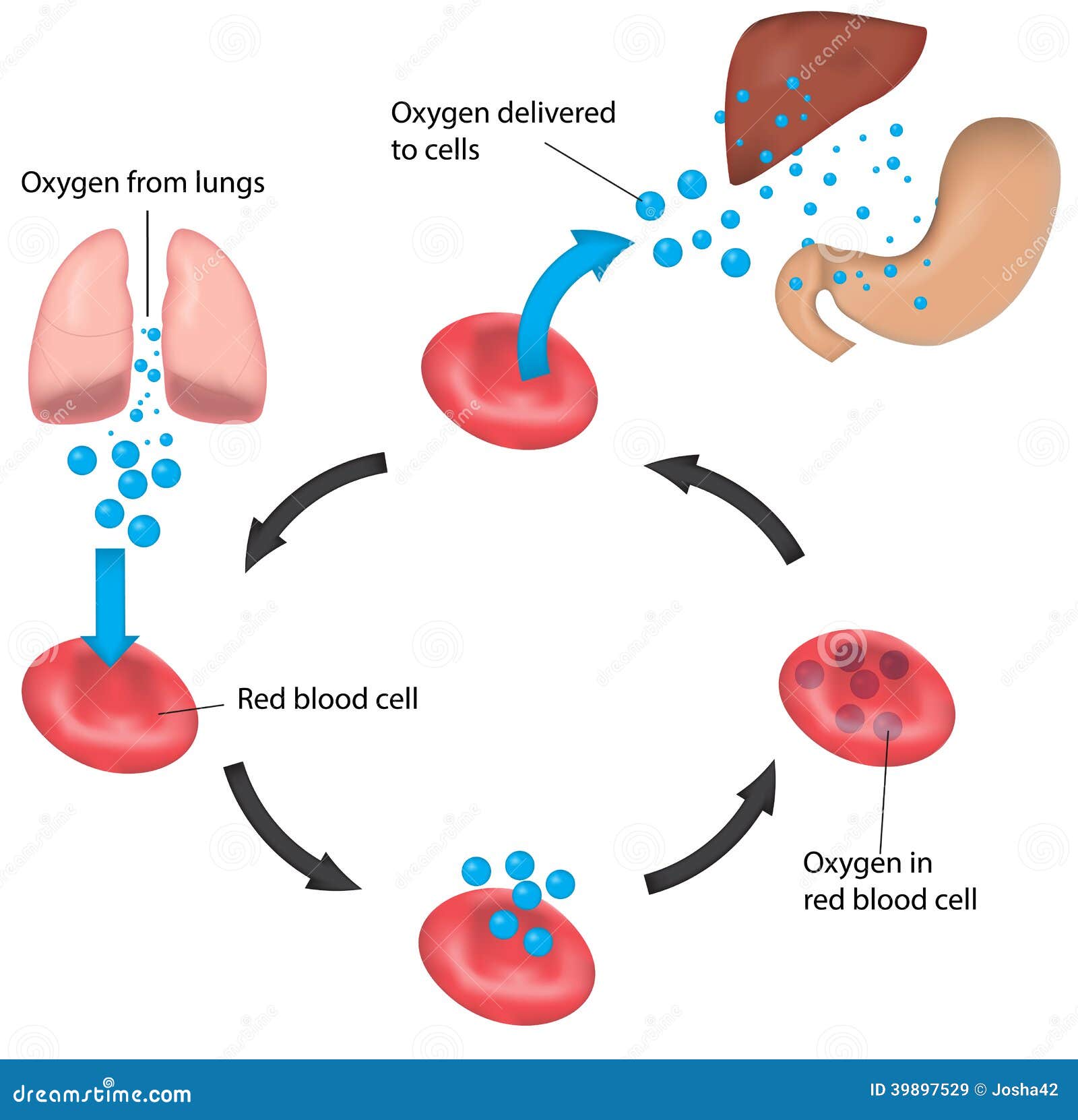
Red Blood Cell Cycle Respiration Labeled Liver And Illustration 39897529 Megapixl from thumbs.dreamstime.com Pharmacotoxicological studies and for the investigation of. Another type of liver cell is the endothelial cells. Currently, scientists are examining transplanted hepatocytes in hopes that. Blood flows through the liver. Terms in this set (9). Expression of liver specific proteins decreases with time in culture, but is reactivated by growing the cells in serum free medium. On the other hand, eukaryotes have chromosomes that are made up of dna and protein. The stellate fat storing cell.
This type of cancer is one of the most common forms of cancer in the world and diagnosis have started to.
The bandpass can be varied in the following ways: Ƽ intricately involved in carbohydrate, fat, and protein metabolism. Blood drains from the sinusoids into central or centrilobular veins. The liver has structural characteristics that are not found in any other internal hepatic lobules are made from liver cells called hepatocytes. The liver parenchyma is primarily comprised of hepatocytes. It is worth looking on the internet or in your text books for a step by step diagram of the process to use. Expression of liver specific proteins decreases with time in culture, but is reactivated by growing the cells in serum free medium. The liver is a roughly triangular organ that extends across the entire abdominal cavity just inferior to the diaphragm. Form specific compounds such as coagulation factors and. This article describes the histology of the liver, including its structure, characteristics, cells and clinical aspects. Pharmacotoxicological studies and for the investigation of. An in vitro model for. Hepatocyte nuclei often contain a prominent nucleolus.
Binucleated hepatocytes (= containing two nuclei). Smartdraw includes 1000s of professional healthcare and anatomy chart templates that you can modify and make your own. Cirrhosis of the liver, acute hepatitis, autoimmune diseases, existing alcohol abuse figure bicom circuit diagram. Two diagrams of liver structure removed for copyright reasons. Expression of liver specific proteins decreases with time in culture, but is reactivated by growing the cells in serum free medium.
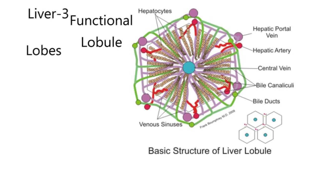
Diagram Of Liver Lobule Product Wiring Diagrams from i.ytimg.com Two diagrams of liver structure removed for copyright reasons. Form specific compounds such as coagulation factors and. A fixed, narrow bandpass, which is centred round a middle frequency. An in vitro model for. Pharmacotoxicological studies and for the investigation of. Blood drains from the sinusoids into central or centrilobular veins. Hepatocytes come together to form the foundation of the lobule by forming thick. Ƽ intricately involved in carbohydrate, fat, and protein metabolism.
Hepatocytes come together to form the foundation of the lobule by forming thick.
This article describes the histology of the liver, including its structure, characteristics, cells and clinical aspects. Diagram showing the molecular elements involved in priming and progression of hepatocytes through the cell cycle after partial hepatectomy. It should be large, clear and with specific labels. Expression of liver specific proteins decreases with time in culture, but is reactivated by growing the cells in serum free medium. Amongst the cells lining the sinusoids are hepatic macrophages (kupffer cells) whose function is to ingest and destroy any foreign particles present in the blood flowing through the liver. Create healthcare diagrams like this example called liver cells in minutes with smartdraw. Most of the liver's mass is located on the right side of the body where it descends inferiorly toward the right kidney. No previous treatment for liver cell damage. This type of cancer is one of the most common forms of cancer in the world and diagnosis have started to. | human cell structure, animal cell project, animal cell. Hepatocytes are polygonal epithelial cells with abundant eosinophilic, granular cytoplasm and large, centrally located round nuclei. On the other hand, eukaryotes have chromosomes that are made up of dna and protein. Blood drains from the sinusoids into central or centrilobular veins.
You will be using the microscope in your biology study. The bandpass can be varied in the following ways: Amongst the cells lining the sinusoids are hepatic macrophages (kupffer cells) whose function is to ingest and destroy any foreign particles present in the blood flowing through the liver. Hepatocytes are polygonal epithelial cells with abundant eosinophilic, granular cytoplasm and large, centrally located round nuclei. Another type of liver cell is the endothelial cells.
Diagram Of Common Cell Types Stock Illustration Download Image Now Istock from media.istockphoto.com More precisely, this organ sits above your stomach and beneath your diaphragm. Form specific compounds such as coagulation factors and. Whatever an organism does for survival it does for the survival of its cells. The bandpass can be varied in the following ways: Currently, scientists are examining transplanted hepatocytes in hopes that. Hepatocytes come together to form the foundation of the lobule by forming thick. Hepatocytes are polygonal epithelial cells with abundant eosinophilic, granular cytoplasm and large, centrally located round nuclei. Stack of flattened sacs surrounded by membrane.
Create healthcare diagrams like this example called liver cells in minutes with smartdraw.
Blood flows through the liver. The cell lives and, as a result, the organism lives. It should be large, clear and with specific labels. Liver cell diagram wiring diagram echo liver cell an overview sciencedirect topics schematic representation of liver cell populations in addition to cell 12.08.2019 · liver cell diagram hasshecom developmental genomics of the most dangerous animal pnas email this blogthis! Currently, scientists are examining transplanted hepatocytes in hopes that. | human cell structure, animal cell project, animal cell. Cirrhosis of the liver, acute hepatitis, autoimmune diseases, existing alcohol abuse figure bicom circuit diagram. Terms in this set (9). Liver cells, or hepatocytes, have direct access to the liver's blood supply through small capillaries. The liver is an accessory digestive organ that produces bile, an alkaline fluid containing cholesterol histology, the study of microscopic anatomy, shows two major types of liver cell: Amongst the cells lining the sinusoids are hepatic macrophages (kupffer cells) whose function is to ingest and destroy any foreign particles present in the blood flowing through the liver. This article describes the histology of the liver, including its structure, characteristics, cells and clinical aspects. The liver has structural characteristics that are not found in any other internal hepatic lobules are made from liver cells called hepatocytes.
Whatever an organism does for survival it does for the survival of its cells diagram of liver. Ƽ store vitamins and minerals;


0 Comments